Lateral eyes
Differences in Eye Size Across Castes and Species
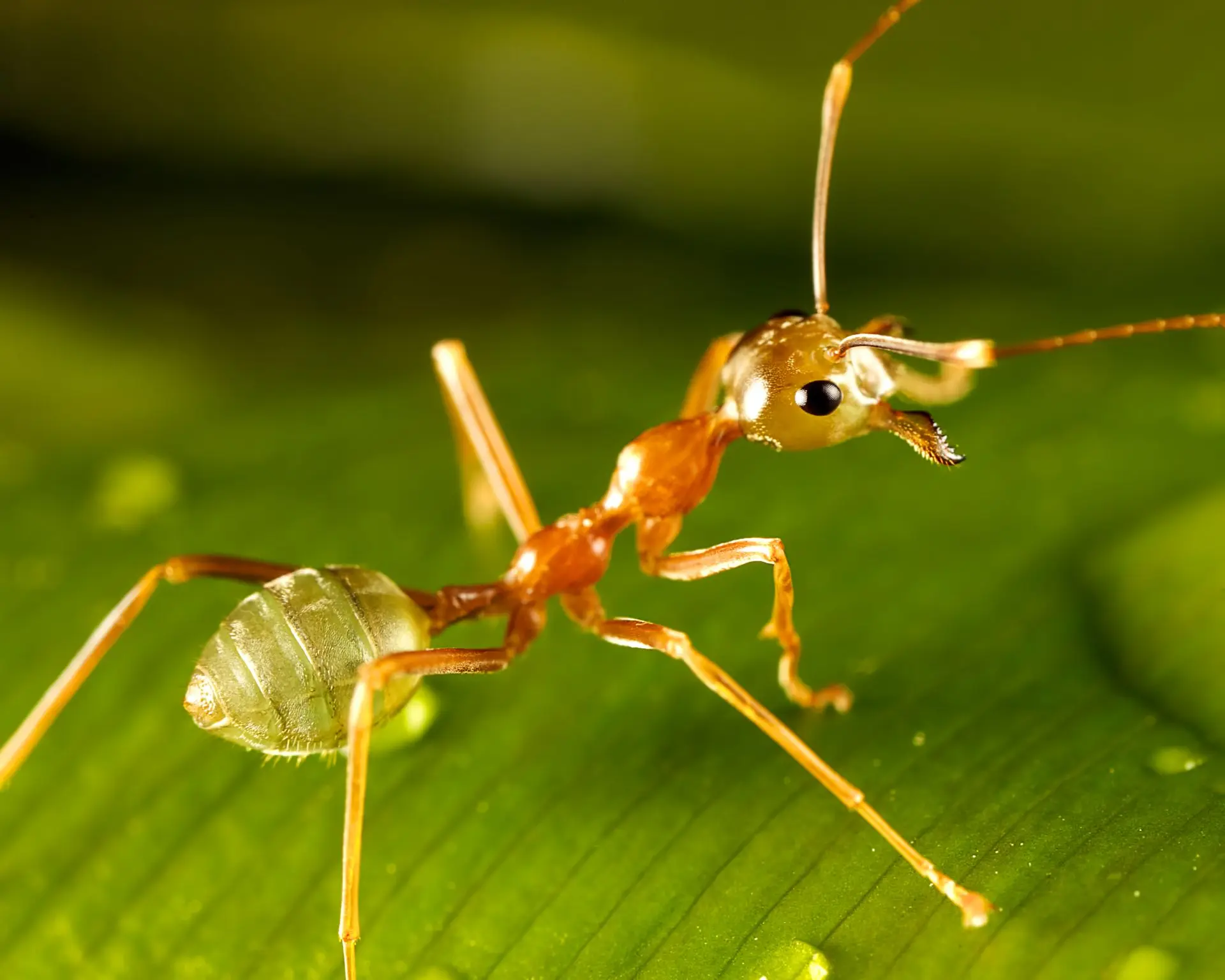
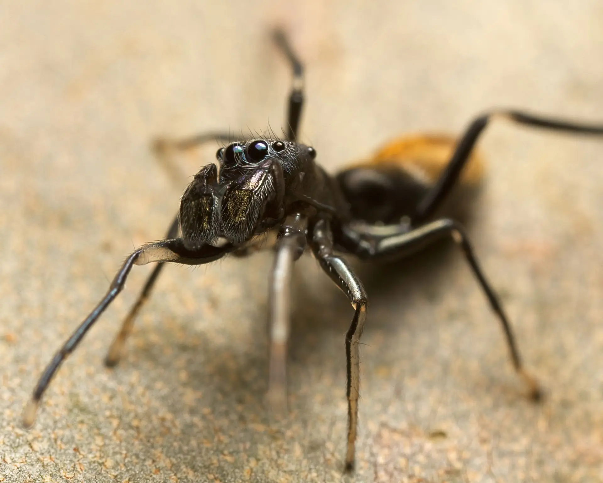
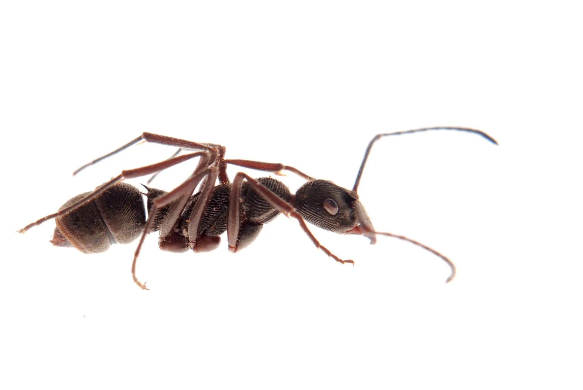
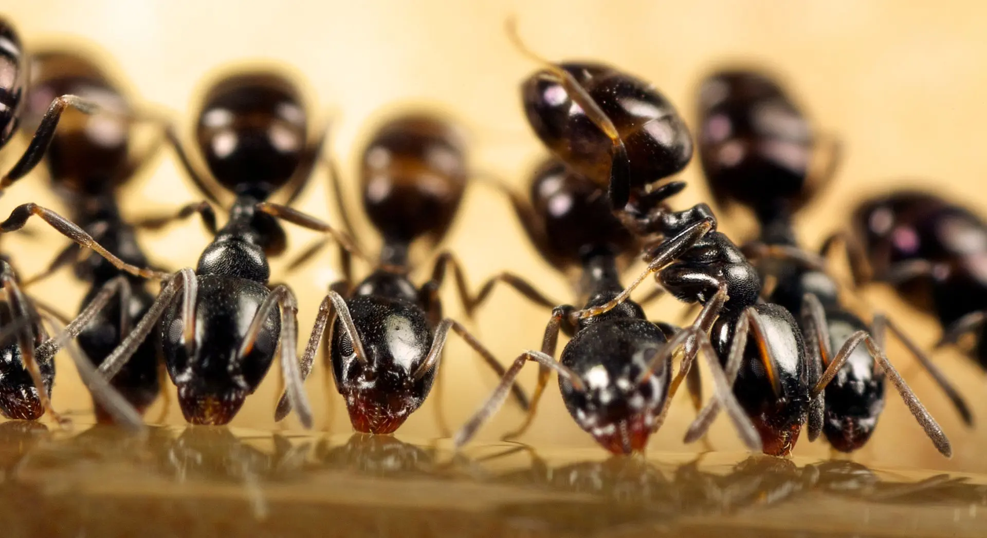
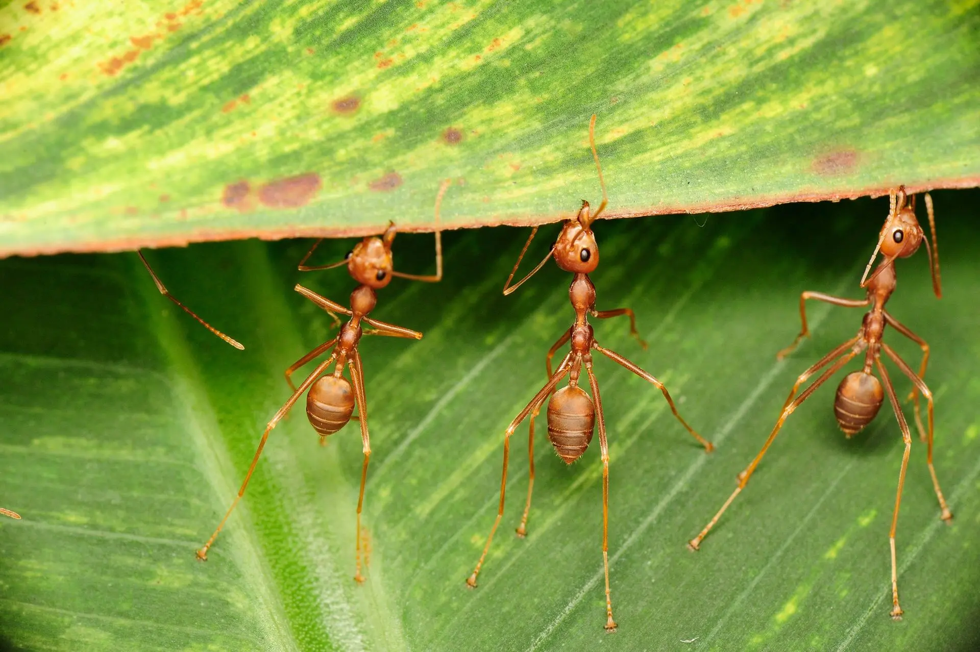
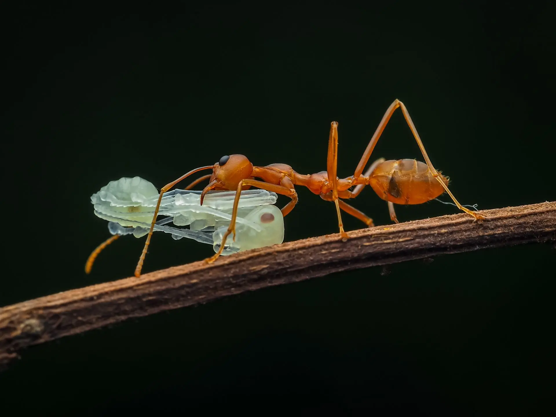
Anatomy of the Compound Eyes
Each of the lateral, or compound, eyes, consists of a closely aggregated mass of sensillae, which are usually called ommatidia and consist of the three elements of the typical protaesthesis : hypodermal cells, which secrete the cornea (facet), gland-cells, which secrete the crystalline cone and nerve-cells known as retinulae. There seem to be no comparative studies on the minute structure of the ommatidia in ants.
It is probable that they differ but little from those of other insects. The great differences in size in the eyes of the different species and castes and their almost universal reduction or degeneration in the workers, induced Forel (1874) to investigate the size and numbers of facets, which, of course, represent the ommatidia. He found the size of the facets varying but little in the different species. The smallest were seen in the male of Cremastogaster sordidula, the largest in the worker of Messor barbarus, but the latter were only twice as broad as the former. The number of facets is extremely variable, from 1 in the worker of Ponera punctatissima, to 1,200 in the male of Formica pratensis. The variation in number of the different castes of the same species is also very striking. Thus, in Tapinoma erraticum, the worker has 100, the female 260, the male 400 ommatidia in each eye; in Formica pratensis, the corresponding numbers are 600, 830 and 1,200, and in Solenopsis fugax 6-9, 200, 400.
Degeneration and Variation of Lateral Eyes in Tropical Ants
A similar study of certain tropical ants (e. g., Eciton, Dorylus, Ponerinae, etc. ) would give an even more surprising range of intraspecific variation in the number of facets. The degeneration of the lateral eyes in the workers has proceeded furthest in the African drivers (Dorylus) and American legionary ants (Eciton). In the former the eyes have disappeared completely, and the same is true of certain species of Eciton, but in many of the latter each eye is reduced to a single facet, which, however, is no longer connected with the brain by an optic nerve.
At any rate, in the specimens of E. schmitti, which I sectioned, I found the vestigial optic nerve reduced to a small thread depending freely from the inner surface of the eye at some distance from the optic lobe.
Caste and Species-Specific Eye Development in Eciton and Aenictus
In some species of Eciton and Aenictus the eyes are represented only by a pair of small, pale dots in the chitinous cranium. While the females of all these genera have no better eyes than the workers, these organs in the males are extremely large and contain many hundreds of ommatidia. It is obvious, of course, that such enormous differences in the size and development of the lateral eyes in different species and in the various castes of the same species must imply corresponding visual differences.